Myoelectric interfaces stimulate and measure the electrical activity of muscle, and may be used to return mobility to the disabled, and study the recruitment of muscles/reflexes. An individual muscle may be composed of multiple neuromuscular compartments, which can differ in both their muscle fiber composition and the torque they exert on the tendon. Thus, for a myoelectric interface to effectively control and observe electro myographic (EMG) activity, the interface must stimulate/measure EMG activity across the entirety of the muscle, match the muscle’s resolution of recruitment, and measure EMG activity with a high signal-to-noise ratio.
Currently, such interfaces are limited in their signal fidelity, spatial resolution, and/or interfacial area. Intramuscular interfaces composed of stiff material scan harm tissue and are restricted their applications; and transcutaneous interfaces possess poor spatial resolution and signal fidelity due to their physical separation from the muscle tissue. DeWeerth's research group developed stretchable electrode array technologies to stimulate and measure EMG activity across a large interfacial area, while stretching with contracting muscle.
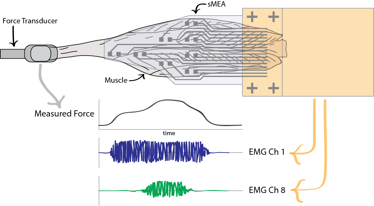
Our stretchable microneedle electrode array (sMEA) interfaces intramuscularly to stimulate and measure EMG activity with high signal fidelity and spatial resolution; and our epimysial microelectrode array was developed to provide similar functionality less invasively. The group assessed the capability of these technologies to measure the regional activity of muscle (Fig) and selectively excite the tissue. Ultimately, our stretchable electrode array technologies may be used to facilitate the development.